

Chronic Hives May Be a Window Into a Larger Inflammatory Disorder
A patient walks into an allergy clinic because of hives. The skin is covered with raised, itching welts that appear, disappear, and migrate unpredictably. Perhaps the eyelids swell or the lips become distorted overnight. Antihistamines may help, but only partially. There is no obvious food trigger, no new detergent, and no single environmental exposure that explains why the disease began. The diagnosis is chronic spontaneous urticaria, or CSU.
The Skin May Be the Messenger, Not the Entire Story
The skin is one of the body's largest immune organs. Like the respiratory and gastrointestinal tracts, it forms a boundary between the body and the outside world. It contains epithelial cells, immune cells, blood vessels, and sensory nerves that communicate continuously with one another. When a hive forms, we see only the final result of that conversation.
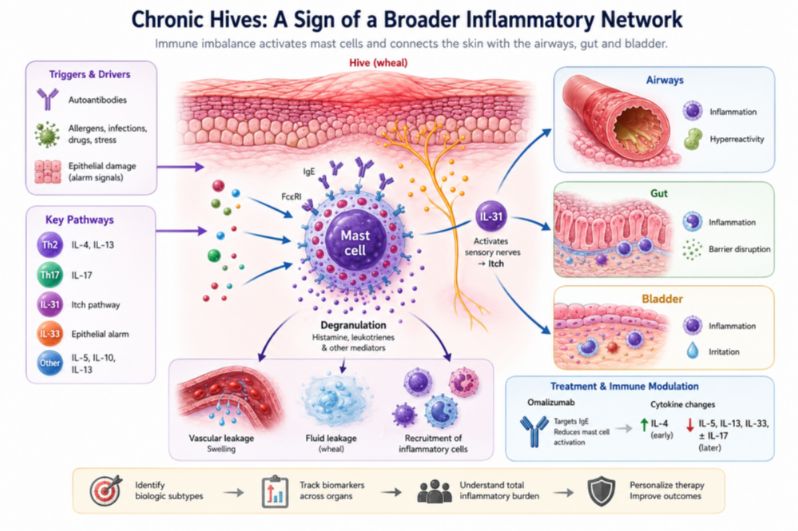
A mast cell releases histamine and other inflammatory mediators. Blood vessels dilate and become temporarily permeable, allowing fluid to move into the surrounding tissue. Sensory nerves are stimulated, and within minutes a raised, intensely itching welt appears. When a similar process occurs deeper in the tissue, angioedema can cause dramatic swelling of the lips, eyelids, hands, feet, or other areas.
Yet mast cells do not act in isolation. They receive signals from IgE antibodies, cytokines, complement pathways, neighboring immune cells, and tissue-derived alarm signals. Their behavior is shaped by the larger inflammatory environment in which they live. From this perspective, the mast cell may be less like a solitary malfunctioning switch and more like a highly responsive participant in a larger immune network.
This broader view may help explain why CSU is so biologically diverse. In some patients, autoantibodies directed against IgE or its high-affinity receptor, FcεRI, appear to contribute to disease. Other patients may have different autoimmune mechanisms, including IgE directed against self-antigens. Still others may have inflammatory patterns that do not fit neatly into either category. The hives may look nearly identical, while the biology beneath them may be quite different.
From a Histamine Disease to an Immune-Network Disease
Histamine remains central to urticaria, and the effectiveness of antihistamines makes that clear. But histamine alone does not explain the full clinical spectrum of CSU. If chronic hives were simply a disorder of excessive histamine, patients might be expected to behave more uniformly. Instead, some achieve excellent control with standard doses of antihistamines, others require intensified therapy, some respond dramatically to omalizumab, some improve only after several months, and others remain difficult to control.
This variability suggests that mast-cell activation may be a common endpoint reached through several different inflammatory pathways. Research on cytokines offers a glimpse into this hidden complexity. IL-17 points toward inflammatory pathways often associated with Th17 immunity and autoimmune disease. IL-31 provides a biological connection between immune activation and the sensory nerves responsible for itch. IL-33 acts as a tissue-associated alarm signal capable of amplifying type 2 inflammation and mast-cell responses. Meanwhile, changes in IL-4, IL-5, and IL-13 suggest that type 2 immunity is also involved, but perhaps in different ways among different patients and at different stages of disease.
CSU may therefore be better understood not as a single broken immune switch, but as a network disorder in which several circuits can malfunction. The skin is simply where that dysfunction becomes visible.
IL-33 and the Language of Tissue Alarm
IL-33 is particularly important to a broader view of inflammatory disease because it belongs to a family of molecules often described as alarmins. These molecules act as biological distress signals. When barrier tissues are injured, irritated, infected, or otherwise stressed, alarmins can alert nearby immune cells and help organize the resulting inflammatory response.In the respiratory tract, epithelial alarm signals are increasingly recognized as important drivers of allergic airway inflammation. In the skin, similar signaling pathways may help connect tissue stress with immune activation. IL-33 can strengthen type 2 inflammatory responses and enhance pathways associated with mast-cell activation.
Studies examining circulating IL-33 in CSU have not been completely consistent. Some have reported increased concentrations, particularly in more severe disease, whereas others have found different patterns. Differences in patient populations, disease phenotypes, laboratory methodology, and the timing of blood collection may partly explain these discrepancies. The inconsistencies also remind us that a single blood measurement may provide only a brief snapshot of a dynamic inflammatory process.
Perhaps more interesting than the absolute concentration of IL-33 is its relationship with other inflammatory signals. IL-33 has been found to correlate with IL-17 and IL-31 in CSU, suggesting potential interaction among tissue alarm pathways, inflammatory T-cell activity, and itch-associated signaling. This is a recurring theme across modern immunology: diseases traditionally classified according to the organ in which symptoms appear may actually involve communication among barrier tissues, immune cells, and the nervous system.
The rash may be local, but the biological conversation that produces it may be considerably broader.
IL-17 and the Autoimmune–Inflammatory Connection
The presence of IL-17-associated inflammation further complicates the traditional picture of chronic hives. IL-17 is closely associated with Th17 cells and with several autoimmune and inflammatory disorders. Once activated, the IL-17 pathway can promote broader inflammatory cascades involving mediators such as TNF-α, IL-1β, and IL-6.
In CSU, higher IL-17 levels have been associated in some studies with greater disease severity. IL-17 has also shown a positive relationship with C-reactive protein, or CRP, a general marker of systemic inflammation. At the same time, higher IL-17 concentrations have been associated with lower circulating basophil counts, another pattern linked in some studies to more active or difficult-to-control CSU.
These findings do not prove that every person with chronic hives has a systemic inflammatory disease. They do, however, challenge the assumption that urticaria must always be understood as an isolated skin disorder. In at least some patients, the visible skin disease may coexist with measurable evidence of a broader inflammatory state.
An important question for future research is whether these biological patterns correspond with inflammatory activity or symptoms in other organ systems. Even more importantly, researchers can ask whether successful control of inflammation in one physiological compartment is followed by measurable improvement elsewhere. That question requires longitudinal observation rather than a single laboratory snapshot.
When the Immune System Creates the Sensation of Itch
IL-31 provides perhaps the clearest example of why strictly organ-based thinking can be limiting. Itching is experienced in the skin, but the sensation requires communication between the immune and nervous systems. IL-31 is produced by immune cells and can interact with receptors connected to sensory nerve pathways, creating a direct mechanism through which immune activation can become a neurological sensation.
This biology may help explain why the severity of itching does not always correspond to the number or size of visible hives. Some patients may have extensive lesions but relatively manageable itching, while others experience severe, relentless discomfort despite having fewer visible wheals. The inflammatory pathway responsible for producing a lesion and the neuroimmune pathway responsible for generating the sensation of itch may overlap without being identical.
A patient does not experience a cytokine level. The patient experiences itching, burning, discomfort, poor sleep, and the exhaustion that follows repeated nights of interrupted rest. The IL-31 story demonstrates how an immune disturbance can cross the conceptual boundary between inflammation and sensation. Other cytokine pathways may prove capable of crossing additional boundaries between organs that medicine has traditionally treated separately.
What Omalizumab Reveals About the Larger Network
Omalizumab provides an unusual window into the biology of CSU. The drug targets free IgE, reducing its availability to interact with the high-affinity IgE receptor on mast cells and basophils. Over time, this changes the sensitivity of these cells to activation.
If CSU were only a simple IgE disorder, the story might end there. However, longitudinal research suggests that omalizumab treatment may be accompanied by changes in a wider cytokine network. During treatment, investigators have observed an increase in IL-4 by the third month, with this change correlating with decreasing disease activity. By six months, IL-5 levels declined significantly, particularly among patients responding to treatment. IL-17 also tended to decrease in some groups, especially among patients with severe disease, although these findings were not uniform across all analyses.
The pattern is intriguing because it suggests that targeting one immune mechanism may gradually reorganize other parts of the inflammatory network. Omalizumab may begin by removing free IgE from circulation, but the downstream consequences can potentially extend through mast cells, basophils, T cells, and their cytokine signals. A drug aimed at one molecular target may gradually influence an entire immune ecosystem.
This possibility could help explain why clinical responses unfold on different timelines. Some patients improve quickly, while others require several months. Early and late responders may show different cytokine trajectories, suggesting that apparently identical hives can emerge from immune systems requiring different biological steps to regain stability.
The broader clinical lesson may extend beyond omalizumab itself. Successful treatment of an inflammatory disease may involve not merely suppressing its final visible symptom, but changing the network that continually recreates that symptom.
Could the Airways and Skin Be Part of the Same Inflammatory Story?
The possibility of biological communication between the airways and skin deserves careful investigation. These organs are usually treated separately, yet immunologically they share important features. Both are barrier systems exposed to the environment. Both contain epithelial cells, mast cells, sensory nerves, and resident immune populations. Both participate in type 2 inflammation, IgE signaling, and tissue alarm pathways.
Asthma, allergic rhinitis, atopic dermatitis, and food allergy are already recognized as biologically interconnected in many patients. The relationship between CSU and airway inflammation is less clearly established and should not be overstated. CSU is not simply another form of asthma, and existing evidence does not prove that airway inflammation causes chronic hives.
The shared biology, however, creates a legitimate and testable research question. If a subgroup of patients with CSU also has active airway inflammation, could inflammatory activity in these compartments move together over time? Could changes in airway control precede, accompany, or predict changes in skin disease? Could common cytokine networks or epithelial alarm pathways help explain why inflammatory diseases cluster in certain patients?
These questions require prospective clinical validation, but they are important because they challenge a deeply rooted habit in medicine: studying each organ independently even when the immune system connecting them is shared.
The Concept of Total Inflammatory Burden
Perhaps the most useful emerging concept is not a single cytokine but the idea of total inflammatory burden. A patient's disease may not be adequately represented by counting hives alone.
One person may have CSU with little evidence of inflammation elsewhere. Another may have hives accompanied by uncontrolled airway inflammation, chronic upper-airway symptoms, gastrointestinal complaints, autoimmune markers, or other signs of immune dysregulation. These patients may share the same diagnostic label while having very different inflammatory phenotypes.
A broader clinical model would therefore assess not only the severity of the hives but also the larger inflammatory landscape. It would examine where else inflammatory activity appears to be present, which immune pathways seem dominant, whether biomarkers change together over time, and whether control of one inflammatory compartment is followed by changes in another.
These questions do not yet have definitive answers, but they are testable. Longitudinal clinical datasets that follow symptoms, pulmonary physiology, inflammatory biomarkers, autoimmune markers, and treatment responses over time may begin to distinguish coincidence from biological connection.
The critical element is temporal sequence. Cross-sectional studies can show that two abnormalities exist at the same time, but they cannot establish how those abnormalities change in relation to one another. If improvement in one inflammatory compartment repeatedly precedes measurable improvement in another, across many patients and over sufficient time, that pattern could reveal relationships that a single laboratory test or office visit cannot detect.
Following Inflammation Across Organs and Across Time
Much of medicine studies disease in snapshots. A cytokine is measured once. A rash is scored during one visit. Pulmonary function is tested on another day. An autoantibody is labeled positive or negative, often without examining how its level changes as the patient's overall inflammatory condition changes.
Inflammatory disease, however, unfolds over time. The more revealing approach may be to follow patients longitudinally and determine whether changes in different organ systems move together in reproducible patterns.
Researchers can ask whether improvement in airway inflammation precedes improvement in chronic hives in a defined subgroup of patients. They can investigate whether IL-17-associated inflammatory patterns identify people with more severe or treatment-resistant disease, whether markers of type 2 inflammation change before symptoms improve, and whether autoimmune markers decline after sustained control of inflammatory activity or instead behave independently.
The ultimate goal would be to identify reproducible inflammatory phenotypes that predict clinical behavior. Such an approach could move medicine away from classifying inflammatory diseases only by the organ in which symptoms are most obvious and toward understanding the biological networks connecting those organs.
A Hive May Be a Biological Clue
A hive is impossible to ignore. It rises visibly from the skin, burns or itches, and then disappears. Because it is so visible, it is tempting to think of the disease as confined to the place where we can see it.The emerging science suggests a more complicated possibility. In some patients, chronic urticaria may represent one visible component of a broader disturbance involving mast cells, IgE pathways, autoimmunity, inflammatory T cells, tissue alarm signals, and neuroimmune communication.
The evidence does not yet justify treating every person with CSU as though they have a systemic inflammatory disease. Nor does it establish that inflammation in one organ necessarily causes disease in another. What the evidence does provide is a strong reason to investigate these connections systematically and longitudinally.
The history of medicine is filled with diseases that were initially classified according to the organ in which symptoms were easiest to observe. As biology became clearer, some of those boundaries dissolved. Chronic spontaneous urticaria may be approaching a similar moment.
The future question may no longer be simply what is causing the rash. It may be whether the skin is allowing us to see part of a larger inflammatory process that has been present all along.
Reference
1. Lin W, Zhou Q, Liu C, Ying M, Xu S. Increased plasma IL-17, IL-31, and IL-33 levels in chronic spontaneous urticaria. Sci Rep. 2017;7(1):17797. Published 2017 Dec 19. doi:10.1038/s41598-017-18187-z
2. Hoşgören-Tekin S, Eyüboğlu İP, Akkiprik M, Giménez-Arnau AM, Salman A. Inflammatory cytokine levels and changes during omalizumab treatment in chronic spontaneous urticaria. Arch Dermatol Res. 2024;316(6):261. Published 2024 May 25. doi:10.1007/s00403-024-02966-6
3. Nguyen D, Deitiker P. Sustained control of recalcitrant chronic spontaneous urticaria after initiation of inflammatory airway diseases treatment: two case reports. J Med Case Rep. 2024;18(1):113. Published 2024 Feb 23. doi:10.1186/s13256-024-04436-z
4. Kolkhir P, Giménez-Arnau AM, Kulthanan K, Peter J, Metz M, Maurer M. Urticaria. Nat Rev Dis Primers. 2022;8(1):61. Published 2022 Sep 15. doi:10.1038/s41572-022-00389-z
5. Maurer M, Rosén K, Hsieh HJ, et al. Omalizumab for the treatment of chronic idiopathic or spontaneous urticaria. N Engl J Med. 2013;368(10):924-935. doi:10.1056/NEJMoa1215372