

Fatal Asthma May Be More Than a Disease of the Airways
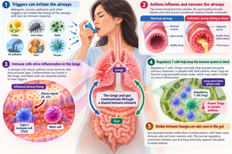
A rare look across human organs suggests that the most dangerous asthma may involve a breakdown of immune regulation connecting the lungs, lymph nodes and gut
Looking Beyond the Lung
The lungs are constantly exposed to foreign material. With every breath, pollen, dust, microbes, pollution and countless microscopic particles encounter an enormous epithelial surface. Most of these exposures do not provoke dangerous inflammation because the immune system must continuously decide what deserves a response and what should be tolerated.
That balance appeared disturbed in people who died from asthma.
All fatal-asthma donors in the study had IgE concentrations above 100 IU/mL, consistent with a strongly allergy-associated, type 2 immune pattern. Their lungs also showed prominent mucus-producing goblet cells and increased populations of immune cells capable of sustaining inflammation.
But some of the most interesting differences were found outside the airways.
Immune abnormalities appeared in lymph nodes associated with both the lungs and gastrointestinal tract. Among younger fatal-asthma donors, populations of naïve T and B cells were reduced while memory populations were increased. In other words, immune tissues in relatively young people had accumulated unusually large populations of cells shaped by previous immune encounters.
This does not mean that asthma literally causes the immune system to age prematurely. The study examined tissues after death and cannot determine when these changes began or what caused them. But it raises the possibility that years of repeated inflammatory stimulation may progressively reshape the immune system that surrounds mucosal organs.
An Immune System That Remembers Too Well
Immune memory is normally one of the body's great advantages. After an infection, memory cells remain behind so that the immune system can react rapidly if the same pathogen returns.
In allergic disease, however, memory can become a liability.
Experiments conducted years before the new human study demonstrated that allergen-specific T cells can take up long-term residence in lung tissue. In a mouse model involving house-dust-mite allergen, these tissue-resident memory cells remained in the lungs after exposure had ended. When the allergen returned, the resident cells alone could help generate airway hyperresponsiveness.
Subsequent work has shown that resident and circulating memory Th2 cells may play different roles during an allergic response. Cells already stationed around the airways can rapidly promote mucus production, eosinophil activation and airway hyperresponsiveness, while circulating memory cells can enter the lungs and amplify inflammation.
The fatal-asthma study now provides an important human counterpart to this experimental work. Increased Th2-like and Th1-like resident-memory populations were found in the lungs of people who died from asthma.
The implication is not simply that inflammation persists. The tissue itself may retain a cellular memory of previous inflammation, leaving the lungs prepared to respond aggressively when another trigger arrives.
When Damaged Airways Sound the Alarm
That memory may become particularly important when the epithelial barrier is injured.
Cells lining the respiratory tract do more than form a physical wall. When damaged by allergens, viruses, pollutants or other stresses, they release signaling molecules that alert the immune system. One of the best studied is interleukin-33, or IL-33.
IL-33 can activate several components of type 2 immunity. Experimental studies have also shown that signaling through IL-33 and its receptor, ST2, helps establish long-lived Th2 memory cells in the lungs after inhaled allergen exposure. When mice lacked IL-33 or ST2, these resident-memory populations and the associated long-term allergic memory were greatly reduced.
The human fatal-asthma study revealed a potentially important parallel. The lungs of fatal-asthma donors contained increased frequencies of CD4 T cells carrying the IL-33 receptor, particularly among type 2 memory populations. Type 2 innate lymphoid cells, another population responsive to epithelial alarm signals, were also increased.
A plausible biological cycle begins to emerge.
Damage to the airway lining releases alarm signals. Those signals activate inflammatory cells already positioned in the tissue. Inflammation produces additional epithelial stress and mucus, potentially releasing still more alarm signals.
Asthma could therefore become self-reinforcing, not because the immune system simply remains switched on continuously, but because repeated injury leaves behind cells capable of restarting inflammation rapidly.
The Missing Immune Brakes
An accelerator, however, is only half of the story.
Healthy immunity also depends on powerful braking systems. Among the most important are regulatory T cells, or Tregs. These cells help maintain tolerance to harmless substances, restrain excessive immune responses and contribute to restoration of tissue homeostasis.
Their importance in asthma has been suspected for years. Experimental and clinical research suggests that disruption of Treg number, stability or function can allow allergic inflammation to persist. Under some chronically inflammatory conditions, regulatory cells themselves may even acquire altered properties that reduce their ability to maintain tolerance.
The organ-donor study found a particularly striking abnormality: regulatory T cells were reduced in lymph nodes draining the lungs of people with asthma. The reduction was especially apparent in younger donors.
This finding changes the emphasis slightly. Severe asthma may not arise only because inflammatory pathways are unusually powerful. The mechanisms responsible for restraining those pathways may also be weakened.
That distinction could eventually matter therapeutically. Suppressing inflammation and restoring immune tolerance are related goals, but they are not necessarily the same biological process.
The Unexpected Signal from the Gut
Perhaps the most intriguing part of the study appeared far from the airways.
Abnormal immune patterns were also detected in mesenteric lymph nodes, which are closely associated with the gastrointestinal tract. In fatal asthma, relationships between immune-cell populations in these gut-associated lymph nodes and populations in the lungs differed from those seen in people without asthma.
This observation adds to growing interest in the gut-lung axis, the idea that immune events in the gastrointestinal tract can influence immunity in the respiratory system and vice versa.
The connection is biologically plausible. The intestinal and respiratory tracts are both enormous mucosal surfaces exposed continuously to the environment. Immune cells activated in one location can circulate elsewhere, while microbial products and metabolites generated in the intestine can influence immune regulation throughout the body.
Earlier research has suggested that the intestinal microbiome can influence the development of regulatory immune responses, particularly early in life. Regulatory T cells in the intestinal mucosa may help restrain type 2 responses, while microbial metabolites can influence immune-cell development and function.
The new human study moves the discussion beyond the microbiome alone. It suggests that the lymphoid tissues associated with the gut and lungs may participate in a larger network whose normal regulatory relationships become altered in asthma.
That does not mean that asthma begins in the gut, nor does the study demonstrate that treating gastrointestinal inflammation will control severe asthma. The researchers observed associations among immune populations, not the direction of causality.
But the findings make it increasingly difficult to view each mucosal organ as an immunological island.
Asthma as a Disease of Barrier Immunity
The broader idea emerging from this research is that asthma may need to be understood partly as a disorder of barrier immunity.
The lungs and gastrointestinal tract share a difficult biological task. They must tolerate enormous numbers of harmless environmental exposures while remaining capable of mounting powerful responses against genuine threats.
That requires coordination among epithelial cells, innate immune cells, T cells, B cells, lymph nodes and regulatory mechanisms.
In fatal asthma, several parts of this system appeared altered simultaneously: memory T and B cells accumulated in mucosal lymph nodes, regulatory T cells were reduced around the lungs, inflammatory resident-memory cells accumulated within lung tissue, IL-33-responsive cells were increased and normal relationships between gut-associated and lung-associated immunity were disrupted.
Viewed together, these findings suggest a disease in which the immune system may become progressively better at remembering inflammatory encounters while becoming less effective at restoring tolerance afterward.
Why This Does Not Mean Every Case of Asthma Is Systemic
The findings should be interpreted cautiously. The fatal-asthma group was small, as would be expected for a study requiring human organs from people who died during an asthma attack. The donors also differed in age, sex, ethnicity, disease severity and medication exposure. More than half of fatal-asthma donors had received corticosteroids, which themselves can influence immune populations.
Most importantly, these tissues represent the endpoint of disease. They cannot reveal whether abnormal immune networks existed years earlier and contributed to severe asthma, or whether severe and repeated inflammation gradually produced the abnormalities that were observed.
The fatal cases were also strongly associated with type 2 immunity and elevated IgE. Asthma is biologically heterogeneous, and the same immune architecture should not automatically be assumed in every person with asthma.
The study therefore does not establish a new clinical test or treatment. It provides something more fundamental: a map showing where investigators may need to look.
Treating the Airways May Be Only Part of the Future
Modern asthma therapy has already moved far beyond simply relaxing airway muscle. Inhaled corticosteroids suppress local inflammation, while biologic medicines can target IgE, eosinophils, IL-4 and IL-13 signaling, and epithelial alarm pathways such as TSLP.
The new findings raise another possibility. Future therapies may eventually be evaluated not only by how effectively they suppress inflammatory cells but by whether healthy immune regulation can be restored.
Could regulatory T-cell function recover when chronic airway inflammation is controlled? Can abnormal tissue-resident memory populations diminish? Does repairing the epithelial barrier reduce the signals that repeatedly awaken inflammatory memory? And can normal communication among mucosal immune tissues be re-established?
Those questions remain unanswered.
But they point toward a different way of thinking about chronic asthma. An asthma attack may be the dramatic event that is seen in the lungs, while the biological conditions that permit repeated attacks may have been developing across a much larger immune network.
The airways may be where the crisis becomes visible. The disease that prepares the ground for that crisis may extend well beyond them.
Reference
1. Lee YA, Wells SB, Yee DC, et al. Dysregulated tissue immunity in human fatal asthma. Nat Commun. Published September 5, 2026. doi:10.1038/s41467-026-77256-y.
2. Hondowicz BD, An D, Schenkel JM, et al. Interleukin-2-Dependent Allergen-Specific Tissue-Resident Memory Cells Drive Asthma. Immunity. 2016;44(1):155-166. doi:10.1016/j.immuni.2015.11.004
3. Rahimi RA, Nepal K, Cetinbas M, Sadreyev RI, Luster AD. Distinct functions of tissue-resident and circulating memory Th2 cells in allergic airway disease. J Exp Med. 2020;217(9):e20190865. doi:10.1084/jem.20190865
4. Kobayashi T, Iijima K, Matsumoto K, Lama JK, Kita H. Lung-resident CD69+ST2+ TH2 cells mediate long-term type 2 memory to inhaled antigen in mice. J Allergy Clin Immunol. 2023;152(1):167-181.e6. doi:10.1016/j.jaci.2023.01.016
5. Jheng MJ, Kita H. Control of Asthma and Allergy by Regulatory T Cells. Int Arch Allergy Immunol. 2025;186(1):87-102. doi:10.1159/000540407