

Have We Been Treating Inflammation Too Far Downstream?
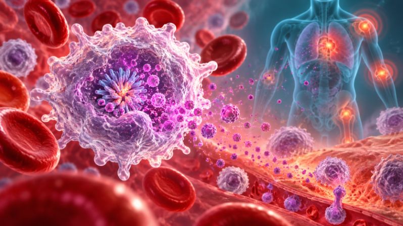
Modern medicine has become increasingly good at blocking the molecules that appear near the end of an inflammatory cascade. Drugs can neutralize cytokines, inhibit enzymes and suppress immune pathways after inflammation is already underway. But a growing body of research is raising a more fundamental question: what if some chronic inflammatory diseases are being treated too far downstream?
The Inflammasome Is Built to Detect Danger
Resistin May Prime the Immune System Before Inflammation Erupts
A Second Pathway May Flip the Inflammatory Switch
Resistin’s role appears to extend beyond priming. The second stage involves Bruton’s tyrosine kinase, or BTK, a signaling enzyme best known for its role in B cells but also active in innate immune pathways. Previous work has shown that BTK can chemically modify NLRP3 by adding phosphate groups to several tyrosine residues. These molecular changes appear to help NLRP3 reorganize into a form capable of assembling the inflammasome complex.
In the new study, resistin was found to interact with BTK and increase its activation. Activated BTK was then associated with phosphorylation of NLRP3, activation of caspase-1 and the subsequent release of mature IL-1β and IL-18. When BTK was inhibited with ibrutinib, inflammasome activity fell and secretion of these inflammatory cytokines decreased. Similar suppression occurred when resistin itself was blocked. The resulting pathway suggests that resistin may operate as more than an inflammatory marker. It may be part of the machinery that transforms extracellular danger signals into full intracellular inflammasome activation.
That creates an intriguing therapeutic possibility. If resistin helps both prepare the inflammasome and activate it, then targeting resistin could theoretically interrupt inflammation at two checkpoints at once. Such an approach would differ from therapies aimed only at NLRP3 or individual cytokines released farther downstream.
Pulmonary Hypertension Offers a Window Into How Inflammation Becomes Structural Disease
The investigators examined this pathway in the context of pulmonary hypertension, a group of disorders characterized by abnormally high pressure in the blood vessels of the lungs. Although the causes of pulmonary hypertension vary, many forms eventually produce similar structural changes. The pulmonary arteries become thicker and narrower, resistance to blood flow rises and the right side of the heart must work progressively harder to pump blood through the lungs. Over time, this increased workload can contribute to right-sided heart failure.
Inflammation has increasingly been implicated in this vascular remodeling. Macrophages can accumulate around pulmonary blood vessels and release cytokines that influence the smooth muscle cells within the vessel wall. Those smooth muscle cells can begin to migrate and proliferate, contributing to thickening of the artery and further narrowing of the vascular channel. In the Johns Hopkins study, fluid collected from resistin-stimulated macrophages activated the growth-related AKT and ERK signaling pathways in human pulmonary vascular smooth muscle cells and increased their proliferation.
The effect was reduced when resistin, NLRP3, IL-1β or IL-18 was blocked. This suggests a possible chain of events in which an inflammatory signal originating in macrophages is translated into structural changes in the pulmonary circulation. Resistin may activate macrophages, NLRP3 may drive the release of IL-1β and IL-18, and those cytokines may then encourage nearby vascular cells to multiply and migrate. Over time, repeated immune activation could therefore contribute not merely to inflammation around the vessels but to the physical remodeling of the vessels themselves.
When Inflammation Begins to Rewrite the Tissue
This connection between immune signaling and tissue architecture is one of the more important implications of the study. Inflammation is often described as redness, swelling or immune-cell activation, but chronic inflammation can eventually alter the structure of organs. Blood vessels can thicken, connective tissue can accumulate, epithelial barriers can be disrupted and smooth muscle can expand. Once these changes become established, the disease is no longer only inflammatory, it has become structural.
The resistin-NLRP3 pathway provides one possible molecular bridge between those two stages. Macrophages respond to stress, inflammatory cytokines are released, neighboring cells receive growth and migration signals, and tissue remodeling follows. This kind of sequence may help explain why some chronic inflammatory diseases become progressively harder to reverse. Once inflammation has repeatedly instructed surrounding tissues to change their behavior, simply reducing one downstream cytokine may no longer be enough to restore normal anatomy.
Human Lung Tissue Suggests the Pathway Exists in Disease
The study also examined lung tissue from nine women with pulmonary hypertension, many of whom had disease associated with systemic sclerosis. Nearly 80 percent of cells expressing NLRP3 in these samples were macrophages, and resistin, BTK and NLRP3 were found together within the same cells. These observations support the biological plausibility of the proposed pathway because the relevant proteins were present in human diseased tissue rather than only in laboratory cell cultures.
At the same time, the human evidence remains preliminary. The patient sample was small, and the study was observational. Finding resistin, BTK and NLRP3 in the same cells does not establish that resistin is the dominant cause of pulmonary hypertension, nor does it show that blocking resistin would improve clinical outcomes. Pulmonary hypertension is biologically diverse, and pathways that are important in one subtype may be less important in another. Larger studies will be needed to determine how consistently this inflammatory circuit appears across patients and whether its activity tracks with disease severity or treatment response.
Mouse Experiments Strengthen the Mechanistic Case
Additional evidence came from mice exposed to low oxygen, a common experimental model of pulmonary vascular remodeling. Mice produce a related protein called RELMα, which performs some functions analogous to human resistin. In normal mice exposed to hypoxia, levels of HMGB1, BTK and NLRP3 increased in the lungs. These increases were largely absent in animals genetically engineered to lack RELMα.
Antibody treatment targeting the resistin-related pathway also reduced hypoxia-induced increases in NLRP3 and IL-1β. These findings strengthen the argument that resistin-like signaling can sit upstream of inflammasome activation rather than simply rising as a consequence of inflammation. Still, animal models cannot fully reproduce the complexity of human pulmonary hypertension, and findings in hypoxic mice should be regarded as mechanistic evidence rather than proof that the same intervention will work in patients.
Why Blocking the End of the Pathway May Not Be Enough
NLRP3 has already become an attractive drug target because of its involvement in inflammatory and metabolic diseases. Experimental compounds such as MCC950 can interfere with inflammasome activation and reduce downstream cytokine production. The new findings, however, highlight a possible limitation of targeting only NLRP3 itself.
Even when inflammasome activation was inhibited, resistin could still initiate the earlier HMGB1-dependent priming program. The immune cell could therefore remain in a prepared inflammatory state. Blocking resistin farther upstream could theoretically reduce both the preparatory phase and the subsequent activation phase. This does not mean resistin will necessarily prove to be a superior therapeutic target, but it raises an important strategic question for drug development: should inflammatory disease be interrupted after the alarm is sounding, or should the signals that repeatedly prepare the alarm system be targeted first?
The distinction may become especially relevant in chronic diseases, where inflammatory pathways are activated repeatedly over months or years. A treatment that suppresses one downstream molecule may provide meaningful benefit, but if the upstream network remains active, inflammation may continue to re-emerge through parallel or compensatory pathways.
Anti-Resistin Therapy Is Promising, but Still Preclinical
The researchers tested an antibody against human resistin and found that it reduced NLRP3-related signaling in cultured macrophages. A related antibody also reduced inflammatory responses in mice. These results make resistin an intriguing therapeutic target because blocking it could potentially interrupt several inflammatory signals simultaneously.
But the experiments do not establish anti-resistin therapy as a treatment for pulmonary hypertension or any other human disease. No anti-resistin antibody was tested in patients in this study. The safety of prolonged resistin blockade is unknown, and resistin may have normal physiological functions that have not yet been fully defined. It will also be necessary to determine which diseases, and which patients within those diseases, are actually driven by this pathway.
The same caution applies to BTK inhibitors such as ibrutinib. Although ibrutinib helped demonstrate that BTK participates in the resistin-NLRP3 pathway, it affects many immune functions and is not being established by this research as a treatment for pulmonary hypertension. Mechanistic experiments can reveal where a pathway can be interrupted, but identifying a drug target and identifying a safe clinical therapy are very different steps.
Chronic Inflammation May Be a Problem of Repeated Priming
Perhaps the most provocative implication of the study is that chronic inflammation may not simply reflect an immune system that refuses to turn off. In some diseases, the deeper problem may be that the immune system is being repeatedly prepared to turn itself back on.
Tissue stress could increase resistin-related signaling. Resistin could stimulate HMGB1 and NF-κB, increasing production of inflammasome components. BTK could then help activate NLRP3, leading to release of IL-1β and IL-18. Those cytokines could alter surrounding tissues, producing additional cellular stress and potentially feeding the inflammatory cycle again. What begins as a protective response could gradually become a self-reinforcing loop.
This concept may extend well beyond pulmonary hypertension. NLRP3 activation has been implicated in cardiovascular disease, metabolic disorders, atherosclerosis and several autoinflammatory conditions. Resistin has independently been associated with obesity, insulin resistance and inflammatory disease. Whether resistin serves as an important upstream regulator across these conditions remains uncertain, but the possibility deserves further investigation.
The More Important Question May Be Where to Intervene
Much of modern immunology has been organized around identifying inflammatory molecules and blocking them. That strategy has transformed treatment for many diseases. But inflammation is a network, not a single pathway, and the location at which that network is interrupted may matter as much as the strength of the blockade.
The resistin-NLRP3 findings suggest that therapies aimed at the earliest stages of inflammatory preparation may eventually deserve as much attention as therapies aimed at cytokines produced near the end. If resistin is confirmed as an upstream regulator in human disease, it could become part of a broader shift toward identifying the signals that repeatedly prime immune cells rather than suppressing inflammation only after it is fully established.
For now, anti-resistin therapy remains an experimental possibility. But the biology raises a larger question that may increasingly shape inflammation research: when chronic disease persists despite downstream treatment, perhaps the immune system is not simply staying switched on. Perhaps it is being switched on again and again.
Reference
1. Kariyawasam U, Lam W, Skinner J, Chakrabarti R, Cox A, Hassoun PM, et al. Human resistin is critical to activation of the NLRP3 inflammasome in macrophages. PLoS One. 2026;21(4). doi:10.1371/journal.pone.0337682.
2. Swanson KV, Deng M, Ting JP. The NLRP3 inflammasome: molecular activation and regulation to therapeutics. Nat Rev Immunol. 2019;19(8):477-489. doi:10.1038/s41577-019-0165-0
3. Bittner ZA, Liu X, Mateo Tortola M, et al. BTK operates a phospho-tyrosine switch to regulate NLRP3 inflammasome activity. J Exp Med. 2021;218(11):e20201656. doi:10.1084/jem.20201656
4. Lin Q, Fan C, Skinner JT, et al. RELMα Licenses Macrophages for Damage-Associated Molecular Pattern Activation to Instigate Pulmonary Vascular Remodeling. J Immunol. 2019;203(11):2862-2871. doi:10.4049/jimmunol.1900535
5. Al-Qazazi R, Lima PDA, Prisco SZ, et al. Macrophage-NLRP3 Activation Promotes Right Ventricle Failure in Pulmonary Arterial Hypertension. Am J Respir Crit Care Med. 2022;206(5):608-624. doi:10.1164/rccm.202110-2274OC