

When Hives Aren’t Really Hives: What a Skin Rash May Reveal About the Immune System
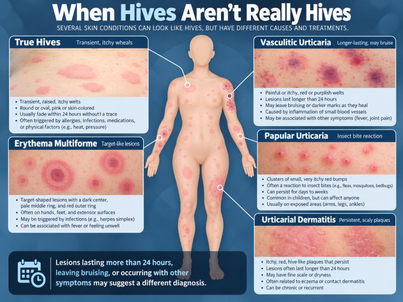
Not every itchy, red welt is caused by an allergy. Some rashes that resemble ordinary hives may be signs of blood vessel inflammation, autoimmune disease, or other immune disorders. The difference often lies in how long the rash lasts, what it leaves behind, and whether other symptoms are present.
The Clue Hidden in How Long a Hive Lasts
Hives are among the most common inflammatory skin conditions. More than 20 percent of people experience acute urticaria at some point in their lives. Chronic urticaria, defined by recurring outbreaks for more than six weeks, affects a smaller proportion of the population and occurs more frequently in women.
Despite its association with allergies, chronic urticaria is often not caused by an identifiable external allergen. Instead, the condition frequently involves abnormal activation of mast cells, specialized immune cells located throughout the skin and other tissues. When activated, these cells release histamine and other inflammatory substances that increase the permeability of nearby blood vessels. Fluid escapes into the surrounding tissue, producing raised, itchy swellings.
In some patients, mast cells are activated by autoimmune mechanisms, including antibodies that interfere with normal immune regulation. In others, physical stimuli such as temperature changes, pressure, or exercise can provoke episodes. Whatever the trigger, ordinary hives share an important characteristic: an individual welt typically disappears within 24 hours without leaving a lasting mark.
This behavior offers a practical diagnostic clue. Although new hives may continue appearing elsewhere on the body, each usually fades relatively quickly. A lesion that remains in precisely the same location for more than a day, becomes painful, or leaves bruising or persistent discoloration may reflect a different inflammatory process.
The 24-hour distinction is not absolute, but it offers a useful starting point for investigating why a seemingly ordinary rash behaves unusually.
When the Problem Lies in the Blood Vessels
Among the more consequential imitators of hives is urticarial vasculitis, a rare condition in which inflammation affects the walls of small blood vessels. At first, the resulting lesions may look like ordinary hives. Over time, however, their differences become more apparent.
Rather than disappearing within hours, the lesions often remain fixed for more than a day. Burning, stinging, or tenderness may accompany the rash, and affected areas can develop purple or brown discoloration as the inflammation subsides. These changes occur because blood vessels themselves are being injured, allowing red blood cells to escape into surrounding tissues.
The process may begin when immune complexes, formed from antibodies bound to other substances, accumulate in vessel walls and activate the complement system, a network of proteins involved in immune defense. This response attracts inflammatory cells, which can damage the vessels rather than merely causing temporary swelling.
For many patients, urticarial vasculitis primarily affects the skin. In others, particularly those with certain abnormalities in complement proteins, inflammation may extend to the kidneys, lungs, joints, or gastrointestinal tract. The condition can also occur alongside autoimmune diseases such as lupus.
Because these complications cannot be identified reliably from appearance alone, a skin biopsy may be necessary to establish the diagnosis. Additional blood and urine testing can help determine whether the inflammation extends beyond the skin.
This distinction has direct consequences for treatment. Antihistamines, which are commonly used for ordinary hives, may provide limited benefit when blood vessel inflammation is the dominant problem. Other medications directed at the underlying immune response may be needed.
The Many Disorders That Borrow the Appearance of Hives
Not all conditions that mimic urticaria are associated with serious systemic disease. Some are common inflammatory skin reactions whose appearance simply overlaps with that of ordinary hives.
Papular urticaria, for example, often develops in children following insect bites. The immune system reacts to substances introduced by mosquitoes, fleas, bedbugs, or other biting insects, producing clusters of intensely itchy bumps that may persist for several days. Unlike typical hives, these lesions do not disappear within a few hours, and repeated exposures can produce recurring outbreaks.
Urticarial dermatitis presents a different variation. Here, features of eczema and hives overlap, creating raised, itchy patches that remain visible and may eventually become dry, scaly, or crusted. These eruptions can be associated with eczema, medications, or contact reactions, making their identification important when conventional treatment for hives fails.
Another imitator, erythema multiforme, produces circular lesions with concentric rings, often described as targets or bullseyes. Some develop a darker center or small blister. The condition is frequently associated with infections, particularly herpes simplex virus, and the lesions generally persist longer than ordinary hives.
Even mast cells themselves can produce a condition that superficially resembles urticaria while arising through a different process. In cutaneous mastocytosis, abnormal numbers of mast cells accumulate in the skin, creating persistent reddish-brown spots. When rubbed, these spots may swell into temporary welts, a characteristic known as Darier's sign. Many childhood cases improve over time, although adult presentations may require further evaluation.
What connects these disorders is their appearance, not necessarily their cause. Similar-looking skin eruptions can emerge from insect hypersensitivity, eczema-related inflammation, infections, or abnormal collections of immune cells.
When the Skin Reflects a Deeper Immune Disorder
Some of the most important hives mimickers arise when immune activity becomes misdirected or poorly regulated.
Bullous pemphigoid, an autoimmune blistering disease most often diagnosed in older adults, illustrates how misleading the earliest symptoms can be. Before recognizable blisters appear, patients may experience weeks or months of intense itching and raised, red patches resembling hives. Eventually, firm, fluid-filled blisters may develop as antibodies damage proteins responsible for holding the skin's layers together.
Because the disease can resemble eczema or urticaria in its early stages, specialized skin testing may be needed before the correct diagnosis becomes clear.
Another group of disorders, known as autoinflammatory syndromes, involves abnormalities in the innate immune system, the body's immediate defense against infection and tissue injury. Instead of producing a localized allergic response, the immune system generates episodes of inflammation that can affect multiple tissues.
Patients may experience recurrent fever, fatigue, joint pain, and eruptions resembling hives. In certain rare inherited conditions, excessive activity of an inflammatory messenger called interleukin-1 contributes to these episodes. Treatments that block this pathway can be effective in selected disorders, whereas antihistamines may do little to control the underlying disease.
A different diagnostic clue is provided by erythema marginatum, a nonitchy rash that forms expanding rings or irregular, winding borders. It may occur in acute rheumatic fever, a complication of group A streptococcal infection, and can also be seen in hereditary angioedema. Its recognition may help direct attention toward conditions that otherwise have little in common with ordinary urticaria.
These examples illustrate a broader principle: the skin is not an isolated organ. Its blood vessels, immune cells, and structural proteins are part of a larger biological network. When inflammation becomes abnormal elsewhere in the body, the skin may provide one of the earliest visible clues.
Why Antihistamines Cannot Treat Every Hive-Like Rash
The growing understanding of these disorders helps explain why a treatment effective for one patient may fail in another whose rash looks almost identical.
For ordinary chronic urticaria, second-generation antihistamines remain the foundation of treatment. When standard doses are insufficient, higher doses may be recommended under medical supervision. Patients with persistent symptoms may benefit from additional targeted medications, including biologic therapies that interfere with pathways involved in mast cell activation.
But the same strategy cannot be applied indiscriminately to every raised, itchy eruption. Urticarial vasculitis may require therapies directed at blood vessel inflammation. Bullous pemphigoid requires treatment of the underlying autoimmune reaction. Certain autoinflammatory syndromes respond to medications targeting specific inflammatory cytokines, while insect-related eruptions require attention to the source of exposure.
The 2025 review is a clinical synthesis rather than a trial demonstrating that one diagnostic strategy improves outcomes. Nevertheless, it highlights an important limitation of treating skin diseases on appearance alone. A lack of response to antihistamines does not necessarily establish an alternative diagnosis, because ordinary chronic urticaria can also be difficult to control. Persistent symptoms, however, make careful reassessment particularly important.
What the Skin Can Tell Us, and What It Cannot
For someone experiencing recurring hives, the most useful observations may be those made between medical appointments. Whether an individual lesion disappears overnight, remains fixed for several days, or leaves discoloration can help distinguish temporary swelling from deeper inflammatory injury. Photographs taken at different times may preserve details that are no longer visible during an examination.
Other symptoms also matter. Unexplained fever, persistent joint pain, unusual bruising, or symptoms suggesting kidney or lung involvement may justify additional investigation. When these features are absent and the lesions behave like typical hives, extensive testing is often unnecessary. When unusual findings are present, a targeted evaluation, sometimes including a skin biopsy, may reveal a different diagnosis.
Sudden throat or tongue swelling, breathing difficulty, or faintness associated with hives requires emergency care because these symptoms may indicate anaphylaxis, a potentially life-threatening allergic reaction.
A Rash Is a Clue, Not a Diagnosis
The central question raised by the review is deceptively simple: When two rashes look the same, are they necessarily caused by the same disease?
Increasingly, the answer from dermatology and immunology is no. Similar skin changes can result from temporary mast cell activation, inflammation of blood vessels, autoimmune injury, or dysregulation of inflammatory signaling. Although these processes can produce overlapping appearances, their clinical consequences and treatment requirements may differ substantially.
For most people, hives remain a manageable condition rather than evidence of serious underlying illness. But when lesions persist, change character, or occur alongside unexplained symptoms, their behavior may reveal information that their appearance alone cannot.
The distinction underscores an essential lesson in medicine: the most visible symptom is not always the disease itself. Sometimes it is the first sign of a more complicated process taking place beneath the skin.
Reference
1. Rojo-Gutierrez MI, Moncayo-Coello CV, Macias Weinmann A, et al. Urticaria and other mimickers of urticaria. Front Allergy. 2025;5:1522749. doi:10.3389/falgy.2024.1522749
2. Zuberbier T, Ansari ZA, Abdul Latiff AH, et al. The international guideline for the definition, classification, diagnosis and management of urticaria. Allergy. 2026;81(8):2582-2632. doi:10.1111/all.70210
3. Matos AL, Figueiredo C, Gonçalo M. Differential diagnosis of urticarial lesions. Front Allergy. 2022;3:808543. doi:10.3389/falgy.2022.808543
4. Kolkhir P, Grakhova M, Bonnekoh H, Krause K, Maurer M. Treatment of urticarial vasculitis: a systematic review. J Allergy Clin Immunol. 2019;143(2):458-466. doi:10.1016/j.jaci.2018.09.007
5. Borradori L, Van Beek N, Feliciani C, et al. Updated S2 K guidelines for the management of bullous pemphigoid initiated by the European Academy of Dermatology and Venereology (EADV). J Eur Acad Dermatol Venereol. 2022;36(10):1689-1704. doi:10.1111/jdv.18220