

Can Autoimmunity Be Reversed?
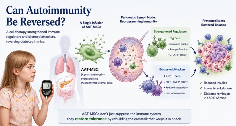
An experimental cell therapy reversed newly developed type 1 diabetes in mice by strengthening the immune system’s peacekeepers and persuading its attackers to stand down
Inside the pancreas, an immune war can unfold almost silently. For months or years, immune cells gather around tiny islands of tissue called the islets of Langerhans. Within them are the β cells that continuously monitor blood sugar and release insulin. At first, the attack may be sporadic. Then immune cells breach the islets in increasing numbers. Cytotoxic T cells recognize β cells as targets and begin destroying them.
Eventually, the pancreas can no longer produce enough insulin to control blood glucose. Type 1 diabetes has arrived. Medicine can replace the missing insulin with extraordinary precision. Continuous glucose monitors can measure sugar levels around the clock. Pumps can deliver insulin automatically. Computer algorithms can connect the two systems, creating something approaching an artificial pancreas.
But the original biological mistake remains unresolved. The immune system still believes that part of the body is the enemy. A study published in Molecular Therapy in 2026 suggests a radically different approach. Instead of broadly suppressing immunity or simply replacing what damaged organs can no longer produce, researchers engineered living cells to intervene in the immune conversation itself.
In mice with newly developed autoimmune diabetes, a single infusion of these modified cells sent more than half of the animals into sustained remission. The treatment appeared to accomplish something immunologists have long sought: it strengthened the cells responsible for maintaining immune tolerance and, through them, restrained the cytotoxic T cells attacking the pancreas.
The result raises a question extending far beyond diabetes. What if some autoimmune diseases are not permanently locked into destruction? What if the immune system can be taught to stop?
The Problem Is Not an Immune System That Is Too Strong
Autoimmune diseases are often described as disorders of an “overactive” immune system. The description is convenient, but incomplete. A healthy immune system must be extraordinarily aggressive. Every day it encounters viruses, bacteria and other threats. It must detect infected cells, mobilize inflammatory signals and, when necessary, destroy parts of the body to protect the whole.
The greater challenge is restraint. The immune system must distinguish a dangerous cell from an innocent one. It must know when inflammation is useful and when it has become destructive. And after an immune response has done its job, the system must know how to stop.
This delicate balance is maintained by an elaborate network of molecular checkpoints and regulatory cells. Among the most important are regulatory T cells, commonly called Tregs.
Tregs are the immune system’s peacekeepers. They patrol the boundary between necessary defense and self-destruction. They suppress inappropriate immune activation, quiet inflammatory signals and help preserve tolerance to the body's own tissues.
In type 1 diabetes, this system of restraint becomes inadequate. Cytotoxic CD8+ T cells and other components of the immune system participate in the destruction of pancreatic β cells. The problem is not simply that inflammatory cells exist. It is that the balance between attack and regulation has shifted.
Researchers have therefore begun asking a different question. Instead of continually suppressing the attackers, could medicine strengthen the cells responsible for keeping them under control?
Building a Living Anti-inflammatory Therapy
The research team led by Wei and colleagues began with mesenchymal stromal cells, or MSCs. These cells have attracted scientific interest because they can influence inflammation and immune-cell behavior. Their therapeutic effects appear to depend less on transforming into replacement tissue than on the signals they release and the interactions they have with surrounding cells.
The researchers then added another component: alpha-1 antitrypsin, or AAT.AAT is best known as a protective protein that shields tissues from enzymes released during inflammation. But its biological effects extend beyond simple enzyme inhibition. AAT has anti-inflammatory and tissue-protective properties that have made it an intriguing candidate for immune-mediated diseases.
The researchers genetically engineered MSCs to produce increased amounts of AAT. The resulting cells, AAT-MSCs, were, in effect, living biological delivery systems. They combined the natural immune-modulating properties of stromal cells with the anti-inflammatory and protective actions of AAT.
The team tested them in non-obese diabetic mice, a widely used model of spontaneous autoimmune diabetes. Crucially, the researchers did not treat the animals before disease appeared. The mice had already developed diabetes.Each received a single intravenous infusion of one million AAT-producing MSCs.
Five weeks later, 11 of 18 treated animals, 61 percent, were diabetes-free. At 10 weeks, approximately 54 percent remained in remission. Among untreated mice, none did. For an experimental autoimmune therapy, the distinction is critical. Preventing disease in a susceptible animal is one challenge. Reversing established disease is another. Something in the immune system had changed.
A Pancreas Under Siege
Under the microscope, the difference was visible. In untreated animals, immune cells had heavily invaded many pancreatic islets. Roughly half showed severe infiltration affecting more than three quarters of the islet. After treatment, severe infiltration fell substantially. At the same time, the proportion of nearly untouched islets increased dramatically.The immune siege was retreating.
The obvious explanation might have been that the engineered cells were simply suppressing inflammation everywhere. But when the researchers examined the immune system more closely, they found something more interesting.The therapy appeared to be rebuilding regulatory control.
Treated animals had increased populations of regulatory T cells carrying molecular features associated with stronger suppressive function. More of these cells expressed CTLA-4 and Helios, molecules associated with immune regulation and Treg stability. And when tested in laboratory experiments, Tregs from treated animals were more effective at suppressing the proliferation of other T cells.
The therapy had not merely produced more peacekeepers. It appeared to have produced better ones.
The Education of a Regulatory T Cell
The researchers used single-cell RNA sequencing and computational analysis to examine how the immune ecosystem changed after treatment. What emerged was a picture of an immune system whose cells were communicating differently.
In pancreatic lymph nodes, the immune staging grounds closely connected to the pancreas, the number of predicted cell-to-cell interactions increased after treatment. Communication networks also expanded within the pancreatic islets themselves. More communication does not necessarily mean more inflammation. The identity of the messenger matters.
After treatment, regulatory T cells became more influential participants in the immune network. Signaling pathways involving important regulatory molecules such as IL-2 and TGF-β were altered in ways consistent with stronger immune restraint. The investigators described the process as a form of Treg “education.”
That phrase captures one of the study’s most intriguing ideas. The engineered stromal cells may not need to remain in the body indefinitely. Their role may be temporary: arrive, alter the inflammatory environment, strengthen regulatory cells and allow those cells to continue reshaping the immune response.
The therapy may act less like a drug that must always be present and more like a teacher whose effects persist through the students. Those students, in turn, appeared capable of influencing the cells attacking the pancreas.
Persuading Killer T Cells to Stand Down
CD8+ T cells are among the immune system’s most powerful weapons. When they recognize a virus-infected cell, they can kill it. This ability is essential for survival. In type 1 diabetes, however, cytotoxic T cells participate in the destruction of insulin-producing β cells.
The AAT-MSC therapy did not simply eliminate these cells. Instead, many began developing characteristics of a biological state known as T-cell exhaustion. The term is deceptive. An exhausted T cell is not tired in the ordinary sense. It has entered a state in which its capacity for aggressive attack is restrained. Such cells often express inhibitory checkpoint molecules and show reduced cytotoxic activity.
In cancer, this phenomenon can be disastrous. Tumors exploit immune exhaustion to escape destruction. Modern checkpoint inhibitor drugs work partly by removing these molecular brakes and restoring T-cell aggression. Autoimmunity presents the opposite problem.
When T cells are attacking the pancreas, joints, nervous system or other healthy tissues, restraint may be precisely what is needed. In the treated mice, more CD8+ T cells expressed TOX, a transcription factor associated with the exhausted state. The cells also showed increased expression of checkpoint molecules including PD-1, TIM-3 and TIGIT.
The researchers then discovered an important connection: Tregs conditioned by the AAT-MSC therapy could help induce this exhausted state in CD8+ T cells. The immune system’s peacekeepers were teaching its soldiers to lower their weapons.
Changing the Conversation, Not Silencing the Room
The broader inflammatory environment changed as well. Signals including interferon-γ, IL-6, TNF-α and IL-1β decreased in pancreatic lymph nodes. These inflammatory molecules can amplify immune activation, recruit additional cells and help sustain cycles of tissue damage. Single-cell analysis revealed changes in genes controlling T-cell activation, inflammatory signaling, cellular movement and responses to antigens.
Taken together, the findings suggest that the therapy did not act through one molecular switch. It altered an ecosystem. That distinction matters because autoimmune disease itself is rarely the result of a single rogue molecule. It emerges from interactions among genetic susceptibility, environmental exposures, tissue injury, antigen presentation, inflammatory signals, immune memory and failures of regulatory restraint.
By the time autoimmune disease becomes clinically visible, these processes may have reinforced one another for years. The therapeutic challenge is therefore not merely to block inflammation. It is to change the system that keeps recreating inflammation.
The Larger Dream of Immune Tolerance
For much of modern medical history, the treatment of autoimmunity has relied on suppression. Corticosteroids suppress inflammation broadly. Other drugs block particular cytokines, eliminate selected immune-cell populations or interfere with pathways necessary for immune activation.
These approaches have transformed the treatment of many diseases. But they generally require continued therapy because the underlying tendency toward autoimmunity remains. The more ambitious goal is immune tolerance. A tolerance-restoring therapy would not simply weaken the immune system. It would restore its ability to recognize that a particular tissue belongs to the body and should not be attacked.
This is one of the central dreams of modern immunology. Type 1 diabetes is an especially difficult test case because stopping the immune attack is only part of the problem. The pancreas must still have enough surviving β cells to recover useful insulin production. If treatment arrives after nearly all β cells are gone, restoring tolerance alone may not be enough.
Timing may therefore be crucial. The mice in the new study had newly developed diabetes, suggesting that some β-cell function remained when treatment began. Whether a similar strategy could work in people with long-standing disease is an entirely different question.
Future therapies may ultimately require combinations: one intervention to restore immune tolerance and another to replace or regenerate lost β cells. But first the attack must stop.
Why the Findings Should Be Viewed with Both Interest and Caution
The results are striking, but they remain findings from mice.The NOD mouse has taught researchers an enormous amount about autoimmune diabetes, but it is not a miniature human. The history of type 1 diabetes research contains many therapies that produced impressive results in mice and far more modest effects in clinical trials.
The study also focused on female NOD mice, which develop diabetes more frequently than males in this model. Whether sex influences the response to AAT-MSC therapy will require further investigation.
There are practical questions as well. Intravenously administered MSCs usually do not take up permanent residence in the body. Many initially localize in the lungs and other organs before disappearing over days or weeks. Yet this apparent limitation creates a fascinating possibility.
Perhaps the engineered cells do not need to survive for years.If they can briefly enter the body, reprogram regulatory T cells and trigger a self-sustaining shift in immune behavior, a short-lived therapy might produce effects lasting far beyond the cells themselves.
That hypothesis remains unproven. But if correct, it could change how scientists think about cellular therapies for autoimmune disease. There are also significant safety and manufacturing challenges. Genetically engineered cells must be produced consistently, tested for potency and molecular stability, and monitored for unexpected long-term effects. Moving from an experimental mouse therapy to a standardized treatment for humans is a long and uncertain process.
The study does not show that type 1 diabetes can now be cured. It shows something more preliminary, but scientifically important: in an animal model of established autoimmune diabetes, immune destruction could be interrupted strongly enough for a substantial proportion of animals to regain control of blood glucose.
Autoimmunity May Be a State, Not a Destiny
The deepest implication of the research may be conceptual. We often imagine autoimmune disease as a switch. Tolerance is present, then it is lost. Once lost, the immune system remains permanently committed to attack. But biology may be more fluid.
The immune system is constantly negotiating between competing forces. Inflammation is opposed by regulation. Cytotoxicity is constrained by inhibitory checkpoints. Tissue damage releases danger signals, while repair processes attempt to restore the barrier. Immune memory can preserve destructive responses, but regulatory networks can also be strengthened.
Disease may emerge when this dynamic system settles into a self-reinforcing state of chronic attack. If so, the central question becomes whether the system can be pushed into another state. The AAT-MSC study offers one experimental answer.
Engineered stromal cells entered the system. Regulatory T cells became more numerous and more suppressive. Communication between immune cells changed. Cytotoxic T cells acquired features of exhaustion. Inflammatory cytokines declined. Immune infiltration of pancreatic islets decreased. And in more than half of treated animals, diabetes went into remission.
None of this guarantees that the same strategy will work in humans. But it points toward a future in which medicine may do more than continuously suppress autoimmune inflammation after it appears. The more ambitious possibility is to repair the failure of tolerance itself, to teach an immune system caught in a cycle of self-destruction how to stop fighting its own body.
Reference
1. Wei H, Gou W, Kim J, et al. Taming autoimmunity: Alpha-1 antitrypsin overexpressing mesenchymal stromal cells promote regulatory T cell crosstalk to reverse diabetes. Mol Ther. 2026;34(7):4181-4199. doi:10.1016/j.ymthe.2026.03.032
2. Sakaguchi S, Yamaguchi T, Nomura T, Ono M. Regulatory T cells and immune tolerance. Cell. 2008;133(5):775-787. doi:10.1016/j.cell.2008.05.009
3. Atkinson MA, Roep BO, Posgai A, Wheeler DCS, Peakman M. The challenge of modulating β-cell autoimmunity in type 1 diabetes. Lancet Diabetes Endocrinol. 2019;7(1):52-64. doi:10.1016/S2213-8587(18)30112-8
4. Bluestone JA, Buckner JH, Fitch M, et al. Type 1 diabetes immunotherapy using polyclonal regulatory T cells. Sci Transl Med. 2015;7(315):315ra189. doi:10.1126/scitranslmed.aad4134
5. Herold KC, Bundy BN, Long SA, et al. An anti-CD3 antibody, teplizumab, in relatives at risk for type 1 diabetes. N Engl J Med. 2019