

How COVID-19 May Nudge the Immune System Toward Autoimmunity
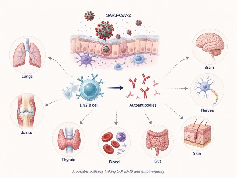
COVID-19 has repeatedly complicated the tidy distinction between infection and autoimmunity. SARS-CoV-2 is a virus, but in some people the immune response it provokes does not remain neatly focused on the pathogen. Antibodies can also emerge against the body’s own proteins, raising the possibility that part of the aftermath of infection may reflect an immune system that has temporarily, or perhaps more persistently, lost some of its ability to distinguish self from nonself.
The Immune System Has to Move Fast, but Speed Has Costs
A B Cell Already Familiar From Lupus
The new study points to a population known as double-negative 2, or DN2, B cells. Their name comes from the absence of two surface markers, IgD and CD27, commonly used to classify different stages of B-cell development.
DN2 cells belong to a broader group known as atypical memory B cells. They express molecules such as CD11c and the transcription factor T-bet, and they appear particularly capable of rapidly transforming into plasmablasts and plasma cells, the specialized factories that secrete antibodies.
These cells were not discovered through COVID-19 research. DN2 cells had previously attracted attention in lupus, where they can become unusually abundant and are associated with the production of autoantibodies. In lupus, they are also especially responsive to molecular pathways that sense nucleic acids.
A similar pattern was seen during SARS-CoV-2 infection. By combining single-cell RNA sequencing, measurements of accessible DNA, plasma protein profiling and extensive autoantibody testing, the researchers found that DN2 cells from people with high autoantibody levels carried molecular features strikingly similar to DN2 cells observed in lupus.
That overlap suggests that infection-associated autoimmunity and classical autoimmune disease may sometimes use the same cellular machinery.
A Faster Route to Antibody Production Circumventing Normal Antibody Training
Many of the immune system’s most refined antibody responses are developed inside temporary structures called germinal centers, which form in lymph nodes and other immune tissues after infection or vaccination. There, B cells repeatedly mutate their antibody genes and compete for survival. Cells producing the most useful antibodies are preferentially selected.
DN2 cells can appear to take a faster path.
During COVID-19, atypical memory B cells have been found to carry relatively low levels of somatic hypermutation, suggesting that many can arise outside the conventional germinal-center process. This faster pathway, known as an extrafollicular response, allows antibody-producing cells to be generated quickly.
Such speed can be beneficial during a rapidly advancing infection. But it may come with a trade-off. A shortened developmental pathway may provide fewer opportunities to remove B cells that recognize the body’s own molecules.
The immune system may therefore face a difficult balance. A pathway that helps generate antibodies rapidly against a dangerous virus can, in susceptible individuals, also increase the chance that self-reactive B cells escape normal restraints.
The Same Sensor That Detects Viral RNA Can Also See Ours
One molecular pathway appears particularly important.
DN2 cells are highly responsive to Toll-like receptor 7, or TLR7, an immune sensor located inside cells. TLR7 detects single-stranded RNA, the genetic material carried by viruses such as SARS-CoV-2.
That makes TLR7 extremely useful during an RNA-virus infection. But it also creates a biological problem because human cells contain RNA as well. TLR7 signaling therefore has to be tightly controlled so that viral RNA is recognized without triggering unnecessary attacks against the body’s own nucleic acids.
In people with high levels of autoantibodies, DN2 cells showed evidence of stronger TLR7-related activity. Additional regulatory changes were also observed in proteins that help control how nucleic-acid-sensing receptors behave.
The researchers then tested the cells directly. When atypical memory B cells isolated from participants were stimulated with a compound that activates TLR7 and the related receptor TLR8, the cells readily developed into antibody-secreting cells. Autoantibodies, including antinuclear antibodies and antibodies against interferon-alpha 2, were produced.
This does not mean that TLR7 activation alone causes autoimmune disease. Instead, the experiments show that this signaling pathway can provide a powerful push toward autoantibody production when the appropriate B-cell population is already present.
Building an Antibody Factory
Two additional molecules help explain how DN2 cells may become so effective at producing antibodies: T-bet and XBP1.T-bet is a transcription factor, a protein that controls the activity of many genes at once. It helps establish the inflammatory identity of DN2 cells and has long been associated with immune responses to viral infection.
XBP1 performs a different job. Antibodies are proteins, and antibody-secreting cells must manufacture extraordinary quantities of them. XBP1 helps reorganize the cell’s internal machinery so that massive protein production becomes possible.
In participants with high autoantibody levels, DN2 cells showed stronger activity involving both regulatory systems. T-bet reinforced the unusual inflammatory state of the cells, whereas XBP1 appeared to prepare them for the transition into high-output antibody factories.
Metabolic changes and signs of increased oxidative stress were also detected, suggesting that these B cells had entered a highly activated physiological state.
Taken together, the findings portray DN2 cells not as passive markers of inflammation but as cells biologically prepared to become potent producers of autoantibodies.
Autoimmunity May Come at the Expense of Antiviral Immunity
The study also uncovered a potentially important relationship between autoimmunity and antiviral protection.
People with higher levels of autoantibodies tended to develop weaker SARS-CoV-2-neutralizing antibody responses during recovery. Higher antinuclear antibody levels during acute illness were also associated with poorer viral-neutralizing capacity months later.
Similar patterns have been reported in earlier longitudinal studies of COVID-19.Several explanations are possible. Immune resources could be diverted toward producing antibodies against self rather than against the virus. Alternatively, the same immune dysregulation that permits autoantibody production may also interfere with the development of an effective antiviral response.
The study cannot distinguish fully between these possibilities. But the observation illustrates an important principle: the strength of an antibody response cannot be judged simply by counting antibodies. The targets those antibodies recognize can be equally important.
An immune response that produces large amounts of antibody is not necessarily an effective one if a substantial portion of that response is misdirected.
Genetics May Help Determine Who Is Vulnerable
The study also raises the possibility that some people may be biologically primed for this type of response.
Could This Help Explain Some Cases of Long COVID?
Long COVID is not a single disorder with a single cause. Persistent viral material, abnormal immune activation, small-vessel and clotting changes, autonomic nervous system dysfunction, latent-virus reactivation and tissue injury have all been proposed as contributing mechanisms.
Autoimmunity is increasingly being investigated as another piece of that puzzle.
Earlier studies have shown that some autoantibodies present during acute COVID-19 are associated with a greater likelihood of persistent symptoms. Other research has documented lasting abnormalities in immune cells and inflammatory proteins in people with Long COVID.
The DN2 study adds another layer by identifying a cellular pathway through which autoantibodies can actually be generated.
That distinction matters. Detecting an autoantibody in blood shows that an unusual immune response has occurred. Identifying the B cells, signaling pathways and transcription factors responsible begins to explain how that response develops.
Still, the evidence stops short of demonstrating that DN2 cells cause Long COVID. Some autoantibodies may be harmless bystanders, whereas others may interfere with immune signaling or damage tissues. Different Long COVID symptoms may also arise from entirely different biological processes.
It is therefore more accurate to say that DN2-driven autoimmunity may contribute to a subset of Long COVID cases, rather than to describe it as a universal explanation.
A Broader Clue About How Infections May Trigger Autoimmunity
The implications may extend beyond COVID-19.For decades, viral infections have been suspected of contributing to autoimmune diseases. Yet the biological route from infection to self-directed immunity has often been difficult to define.
The DN2 pathway provides a plausible bridge.
An RNA virus strongly activates innate sensors such as TLR7. Under intense inflammatory conditions, rapidly responding B cells can expand outside the usual germinal-center training system. T-bet reinforces their inflammatory identity, and XBP1 equips them for massive antibody production. If some of those B cells recognize human proteins, autoantibodies can be produced alongside antiviral antibodies.
Most such responses may disappear as the infection resolves. In genetically or immunologically susceptible people, however, the process may persist long enough to contribute to prolonged inflammation or autoimmune symptoms.
A central question now is what determines which direction the immune system takes.
DN2 cells are not yet a clinical test for Long COVID, and therapies targeting TLR7, T-bet or XBP1 are not established treatments for post-COVID illness. But a cellular mechanism linking antiviral immunity with autoantibody production is becoming clearer.
That mechanism may eventually help explain one of the most puzzling features of viral infections: why the same pathogen can produce a brief illness in one person but leave another with an immune system that continues behaving as though the danger has never fully passed.
Reference
1. Yuan D, Li S, Zhang R, et al. A distinct effector B cell population drives autoantibody production in SARS-CoV-2 infection. Immunity. Published online August 28, 2026. doi:10.1016/j.immuni.2026.08.002.
2. Jenks SA, Cashman KS, Zumaquero E, et al. Distinct Effector B Cells Induced by Unregulated Toll-like Receptor 7 Contribute to Pathogenic Responses in Systemic Lupus Erythematosus. Immunity. 2018;49(4):725-739.e6. doi:10.1016/j.immuni.2018.08.015
3. Su Y, Yuan D, Chen DG, et al. Multiple early factors anticipate post-acute COVID-19 sequelae. Cell. 2022;185(5):881-895.e20. doi:10.1016/j.cell.2022.01.014
4. Woodruff MC, Ramonell RP, Haddad NS, et al. Dysregulated naive B cells and de novo autoreactivity in severe COVID-19. Nature. 2022;611(7934):139-147. doi:10.1038/s41586-022-05273-0
5. Klein J, Wood J, Jaycox JR, et al. Distinguishing features of long COVID identified through immune profiling. Nature. 2023;623(7985):139-148. doi:10.1038/s41586-023-06651-y