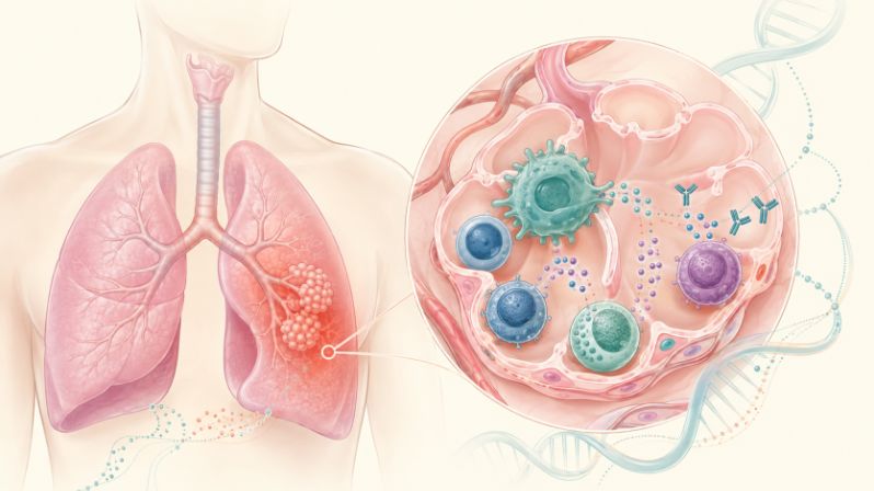
An analysis of more than one million human immune cells suggests that inherited risk for asthma, COPD and autoimmune disease may be expressed in cells that remain inside tissues, where it can be missed by conventional blood tests.
The Immune System Has a Geography
Every breath carries a biological challenge. Viruses, allergens, smoke, dust and chemical particles repeatedly encounter the delicate surfaces of the lungs. Protection is provided partly by immune cells circulating through the blood, but a substantial defensive force remains stationed inside the lung itself.
These tissue-resident immune cells include memory T cells, macrophages, natural killer cells and B cells. Some remember previous infections, some remove damaged cells, and others coordinate inflammation and tissue repair. Their permanent address matters because a cell living in the lung can behave differently from a similar-looking cell traveling through the bloodstream.
A study published in Nature Immunology suggests that this local immune geography may also determine how inherited disease risk is expressed. Genetic variants associated with asthma, chronic obstructive pulmonary disease, or COPD, sarcoidosis, lupus and other autoimmune conditions were connected to changes in gene activity within specific populations of lung-resident immune cells.
The findings do not show that these variants inevitably cause disease. Instead, they provide a possible biological bridge between inherited DNA differences and the inflammatory behavior of immune cells inside an affected organ.
Looking Beyond the Blood
Much of human immunology has been built from blood samples because blood can be collected safely and repeatedly. Yet studying the immune system only through blood is a little like trying to understand a city by watching traffic on its highways. Movement can be seen, but much of what happens within individual neighborhoods remains hidden.
To examine one of those neighborhoods, cancer-free lung tissue was obtained from 120 people undergoing surgery for early-stage lung cancer. More than 1.15 million immune cells were analyzed with single-cell RNA sequencing, which measures the genes active in individual cells. Twenty-nine immune-cell subsets were identified, including multiple groups of resident memory T cells, natural killer cells, B cells, monocytes, macrophages and dendritic cells.
The donors’ genetic variants were then compared with gene activity in each cell population. Nearly 6.9 million common variants were tested. In total, 3,550 genes showed expression differences associated with a person’s genotype. The regulatory variants producing such effects are known as expression quantitative trait loci, or eQTLs.
Many of these genetic effects were shared among related cells, but 894 genes displayed effects restricted to a particular immune-cell type. Another 954 genotype-dependent associations were especially prominent in lung immune cells and had not been apparent in earlier analyses of circulating immune populations.¹ This distinction matters because a disease-linked variant may appear silent in blood while altering gene activity in a cell that has taken up long-term residence in the lung.
How Inherited Risk Could Become Inflammation
Most common disease-associated variants do not break a gene or dramatically change the protein it produces. Many sit in regulatory regions of DNA that help determine when, where and how strongly a gene is activated. They can function like dimmer switches, producing subtle effects that depend on the cell reading the DNA and the conditions surrounding that cell.
Several layers of evidence were combined in the lung study. Disease-associated variants were first mapped to accessible regions of DNA in particular immune cells. These open regions are available to the molecular machinery that controls gene activity. The investigators then asked whether different versions of a variant were associated with higher or lower expression of a nearby gene. Finally, statistical colocalization was used to test whether a disease-risk signal and a gene-expression signal were likely to arise from the same underlying genetic region.
The analysis identified 492 genes whose regulatory signals overlapped with genetic associations for lung, infectious or autoimmune diseases. A possible chain of events can therefore be proposed: an inherited variant changes the regulation of a gene in a particular lung immune cell, the altered gene activity changes how that cell responds to a trigger, and the modified response affects the intensity or duration of inflammation.
This chain remains a hypothesis, not proof that the variants directly cause inflammation. Most participants were not followed from environmental exposure through immune activation to clinical disease. Yet the study narrows the search by identifying the immune cells and molecular pathways in which those causal steps can now be tested.
COPD and Asthma May Share More Genetic Biology Than Expected
COPD is usually associated with cigarette smoke, air pollution and injury to the structural cells of the lungs. These factors remain central to the disease, but the new findings suggest that inherited differences in lung immune cells may also influence susceptibility.
The researchers estimated that gene regulation within lung immune cells mediated approximately 15 to 20 percent of COPD risk attributable to the common genetic variants included in the analysis. The corresponding estimate was about 10 percent when circulating immune cells or mixed, bulk tissue samples were examined.
This finding does not mean that lung immune cells cause 15 to 20 percent of all COPD cases. It is a statistical estimate of the portion of common-variant heritability that may be transmitted through measured changes in gene expression. The estimate is model-dependent and does not include smoking, pollution, occupational exposures, infections or many other factors that influence COPD.
Among the strongest disease associations were those found in CD4-positive and CD8-positive T cells, particularly tissue-resident memory T cells. These long-lived cells remain in the lungs after an immune encounter and can respond rapidly when the same threat returns.
An important signal was found at the chromosome 17q21 region, which has long been associated with asthma. Variants in this region were linked to the activity of ORMDL3, GSDMB and GSDMA in several lung immune-cell populations. One COPD-associated variant was connected to increased ORMDL3 expression in lung-resident T cells. Earlier work had also shown that asthma-risk variants can alter ORMDL3 activity and T-cell function.
Of the 127 genes linked to asthma risk in the new analysis, 72, or 57 percent, were also associated with COPD or impaired lung function. This overlap suggests that some molecular pathways may be shared between the two diseases. However, the authors noted that some COPD datasets may have included people with asthma or asthma-COPD overlap, potentially increasing the apparent similarity.
The Lung May Reflect Systemic Autoimmune Risk
Although the study focused on lung tissue, genetic associations were detected for 32 immune-mediated diseases. A total of 281 genes in lung immune cells showed overlapping signals with systemic autoimmune conditions, including sarcoidosis, systemic lupus erythematosus, rheumatoid arthritis, Sjögren disease and systemic sclerosis.
One of the most notable genes was ZFP57. Its expression was associated with genetic risk for sarcoidosis, an inflammatory disease that frequently affects the lungs, as well as signals from several other autoimmune and lung diseases. When ZFP57 activity was reduced experimentally in laboratory-grown macrophages, the expression of inflammatory cytokine and chemokine genes was altered. This provides preliminary functional evidence that the gene may influence macrophage behavior, although its role in patients remains to be established.
Another gene, IRF5, was linked to risk variants associated with lupus, rheumatoid arthritis and Sjögren disease. A risk-associated variant was connected to increased IRF5 expression in lung myeloid cells and CD4-positive tissue-resident memory T cells. Because IRF5 helps activate inflammatory programs, these cell populations may represent locations where systemic autoimmune risk is expressed.
Disease-linked effects were also found in lung B cells. Variants affecting FAM167A, BLK and TSPAN3 were associated with lupus, rheumatoid arthritis or systemic sclerosis. In systemic sclerosis, genetic effects involving components of the interleukin-12 receptor were separated by cell type, with one receptor subunit affected mainly in resident T cells and another in natural killer cells.
These results do not demonstrate that autoimmune diseases begin in the lungs. Rather, they suggest that genetic programs associated with systemic autoimmunity can operate within immune cells embedded in an affected organ. Similar tissue-resident populations are found in the skin, joints, salivary glands and intestines, but their regulatory patterns will need to be studied directly rather than assumed to be identical.
through a single universal pathway.
Sex Differences in Immunity Are More Complicated Than Gene Activity Alone
Biological sex is associated with substantial differences in susceptibility to autoimmune and infectious diseases. More than 1,700 genes showed sex-biased expression in at least one lung immune-cell population. Genes involved in inflammatory signaling, immune regulation, tissue repair and immune-cell recruitment were among those affected.
However, only 117 genes showed significant differences between males and females in how a genetic variant influenced expression. More than 98 percent of these genes did not also show a simple male-female difference in their overall activity.
This separation suggests that two distinct processes are involved. A gene may be more active in one sex because of hormones, cell composition or environmental influences, while the effect of an inherited variant on that gene may remain similar in both sexes. Conversely, a variant can produce different regulatory effects in males and females even when average gene expression is comparable. The findings do not provide a single genetic explanation for the greater prevalence of many autoimmune diseases in women, but they show why the biology cannot be understood from average gene-expression levels alone.
Important Limitations Remain
The lung samples came from treatment-naive patients with early-stage lung cancer, although tissue was collected from cancer-free areas. Cancer, smoking history, age and other medical conditions could still have influenced the immune cells. The participants were also predominantly older adults, with a median age of 71, so the findings may not fully represent younger populations.
Only lung parenchyma was studied. Immune cells in the airway wall, where much of asthma-related inflammation occurs, may have different genetic programs. Tissue processing also prevented granulocytes, including important populations such as neutrophils and eosinophils, from being adequately analyzed.
Finally, most of the reported relationships were based on statistical colocalization. Laboratory experiments and studies of people with the relevant diseases will be required to determine whether the prioritized variants and genes directly cause harmful immune activity.
Why the Findings Matter
The study reinforces a broader change in immunology: disease risk is increasingly being examined in the cells and tissues where disease actually occurs. A blood sample can reveal much about the immune system, but it may miss genetic effects that appear only after immune cells settle into an organ and adapt to its environment.
No new genetic test or treatment is established by these findings. However, the resulting lung immune-cell atlas may help researchers select more precise drug targets, distinguish protective immune responses from harmful ones and investigate why the same genetic pathway can be associated with asthma, COPD, infection and autoimmunity.
Genetic risk is not destiny. Smoking, allergens, infections, pollution, occupational exposures and other environmental factors remain important determinants of respiratory health. The findings instead suggest that these exposures may be interpreted through immune cells whose responses are partly shaped by inherited genetic differences, and that the most informative place to study those differences may be inside the affected tissue itself.
Reference
1. Schmiedel, B.J., Gonzalez-Colin, C., Fajardo-Rosas, V. et al. Tissue-resident immune cells drive genetic risk in autoimmune and lung diseases. Nat Immunol (2026). doi.org/10.1038/s41590-026-02596-2
2. Li J, Xiao C, Li C, He J. Tissue-resident immune cells: from defining characteristics to roles in diseases. Signal Transduct Target Ther. 2025;10(1):12. Published 2025 Jan 17. doi:10.1038/s41392-024-02050-5
3. Shrine N, Izquierdo AG, Chen J, et al. Author Correction: Multi-ancestry genome-wide association analyses improve resolution of genes and pathways influencing lung function and chronic obstructive pulmonary disease risk. Nat Genet. 2023;55(10):1778-1779. doi:10.1038/s41588-023-01531-7
4. Schmiedel BJ, Seumois G, Samaniego-Castruita D, et al. 17q21 asthma-risk variants switch CTCF binding and regulate IL-2 production by T cells. Nat Commun. 2016;7:13426. Published 2016 Nov 16. doi:10.1038/ncomms13426
5. Allen EK, Randolph AG, Bhangale T, et al. SNP-mediated disruption of CTCF binding at the IFITM3 promoter is associated with risk of severe influenza in humans. Nat Med. 2017;23(8):975-983. doi:10.1038/nm.4370