

The Airway's Early Warning System: How the Lining of Your Lungs Decides Whether You Stay Healthy, or Develop Asthma
For decades, asthma was viewed primarily as a disorder of overactive immune cells. Doctors focused on eosinophils, IgE antibodies, mast cells, and the inflammatory molecules they produced. These immune players were certainly important, but they were not the first to react.
The Lung's Border Patrol
Every day an adult breathes nearly 20,000 liters of air. That air contains oxygen, but it also carries an astonishing assortment of biological and chemical passengers: pollen grains, fungal spores, dust mite proteins, viruses, bacteria, wildfire smoke, diesel exhaust, microscopic plastic particles, and industrial pollutants.
The remarkable challenge facing the lungs is not simply keeping harmful substances out. It is learning what can safely be ignored. If the immune system reacted aggressively to every airborne particle, breathing itself would become impossible.
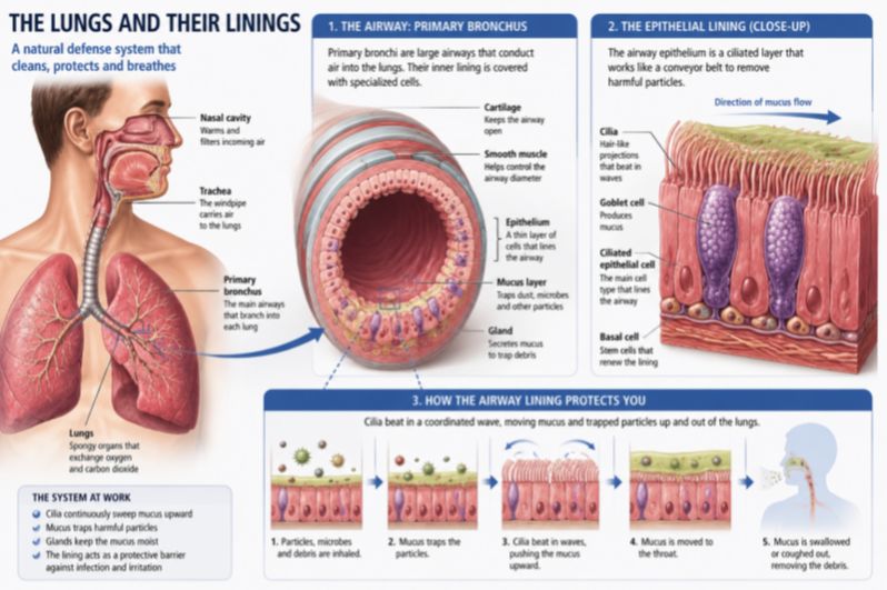
Instead, the airway epithelium functions as an intelligent border patrol. Its tightly joined cells form a physical barrier while simultaneously sampling everything that passes across its surface. Under healthy conditions, these cells encourage immune tolerance, allowing harmless substances to come and go without provoking inflammation.
But barriers can fail. Repeated exposure to allergens, respiratory infections, cigarette smoke, urban air pollution, ozone, and other environmental insults can injure epithelial cells or loosen the microscopic junctions that hold them together. When that happens, the border patrol changes its mission. Instead of maintaining peace, it sends out emergency distress signals.
Three Molecules That Can Change Everything
The first responders are not immune cells. They are the epithelial cells themselves.
TSLP: The Master Dispatcher
Among the earliest alarm signals is TSLP, a molecule that functions almost like an emergency dispatcher. Once released, TSLP activates dendritic cells, the immune system's professional scouts. These scouts travel to nearby lymph nodes, where they educate naïve T cells to become Type 2 helper T cells, the architects of allergic inflammation.
Those newly activated cells begin producing familiar inflammatory molecules such as IL-4, IL-5, and IL-13, which recruit eosinophils, stimulate mucus production, narrow airways, and amplify allergic responses throughout the lungs. Remarkably, scientists have discovered that TSLP exists in two forms. One appears to help maintain healthy epithelial function under normal conditions, while the other is produced primarily during inflammation. It is a reminder that molecules involved in disease often evolved first to protect us.
IL-25: Turning Up the Volume
If TSLP starts the conversation, IL-25 makes sure everyone hears it. Produced by epithelial cells, including recently discovered sensory "tuft cells" scattered throughout the airway, IL-25 powerfully amplifies Type 2 immune responses. Its influence extends beyond inflammation.
IL-25 directly stimulates fibroblasts, the structural cells responsible for producing connective tissue. Over time these cells deposit collagen and other extracellular matrix proteins that gradually thicken airway walls, contributing to the permanent remodeling seen in severe asthma. Asthma, in other words, is not simply inflammation. It is also slow architectural change.
IL-33: The Cellular Distress Beacon
IL-33 may be the most unusual member of the trio. Unlike most cytokines, which are actively secreted, IL-33 normally resides quietly inside the nuclei of healthy epithelial cells. Only when those cells are unexpectedly injured does IL-33 escape into surrounding tissues, acting almost like a distress flare announcing that damage has occurred.
Once released, IL-33 rapidly awakens mast cells, eosinophils, basophils, and a recently discovered family of immune cells called group 2 innate lymphoid cells (ILC2s). These cells require no prior training or antibody recognition. They respond almost immediately, producing enormous quantities of inflammatory cytokines that can transform a local injury into widespread allergic inflammation within hours.
Fortunately, nature built in safeguards.IL-33 quickly becomes chemically oxidized after its release, limiting how long it remains active. The body also produces a soluble "decoy" receptor that captures excess IL-33 before it can continue stimulating immune cells. Even our alarm systems have built-in off switches.
Different Triggers, One Common Language
One of the most fascinating discoveries is that completely different environmental exposures all activate the same epithelial alarm system. Dust mites do it. Cat dander does it. Mold spores do it. Respiratory viruses, including those responsible for the common cold, do it. Wildfire smoke, diesel exhaust, and microscopic air pollutants do it as well.
Although these exposures have little in common biologically, they share one crucial characteristic: they stress or damage the epithelial barrier. The lungs appear less concerned with what caused the injury than with the fact that injury occurred at all. This helps explain why asthma often worsens during viral infections, pollen seasons, episodes of heavy air pollution, or after repeated exposure to cigarette smoke. Each activates the same molecular alarm system from a different direction.
Why Allergies Become So Difficult to Stop
If the body possesses several alarm molecules instead of just one, why hasn't evolution simplified the system? Because redundancy saves lives. If one alarm fails during an infection, others can still summon help. For survival against dangerous microbes, overlapping alarm systems are enormously beneficial.
But in chronic allergic disease, this same redundancy becomes problematic. Animal studies show that blocking a single alarmin often produces only modest improvement because the remaining molecules continue driving inflammation. Blocking all three simultaneously produces far greater reductions in airway inflammation and eosinophil accumulation.
From the immune system's perspective, persistent danger demands persistent vigilance. From the patient's perspective, it means chronic asthma.
A Revolution in Asthma Treatment
For decades asthma therapies focused on the middle or end of the inflammatory cascade. Bronchodilators relaxed airway muscles. Steroids suppressed inflammation. Newer biologics blocked IL-5, IL-4, or IgE after allergic responses were already well underway.
The newest generation of treatments moves much farther upstream. Instead of interrupting the fire after it has spread, scientists are attempting to silence the smoke alarm itself. The first major success is tezepelumab, an antibody that neutralizes TSLP before it can activate the rest of the immune cascade.
Unlike earlier biologic drugs that work primarily in patients with high eosinophil counts, tezepelumab has reduced asthma attacks across a surprisingly broad range of patients, including many whose inflammation does not fit traditional allergic patterns.
Researchers are now exploring therapies aimed at IL-33 and its receptor ST2, with several promising drugs already advancing through clinical trials. Treatments directed against IL-25 remain under active investigation. The field is steadily shifting from treating inflammation to preventing its initiation.
Asthma Begins at the Surface
The growing understanding of epithelial alarmins is changing more than treatment, it is changing how scientists think about asthma itself. Rather than viewing asthma solely as a malfunctioning immune system, researchers increasingly see it as a disease that begins where the body meets the outside world.
The airway epithelium is not simply a protective coating. It is a sophisticated communication network that decides when environmental exposures deserve tolerance and when they warrant alarm. Most of the time, it makes the right decision. But when repeated injury, from allergens, pollution, viruses, tobacco smoke, or other environmental stressors—keeps pulling the alarm, the immune system gradually shifts into a state of chronic vigilance. What began as a protective response slowly evolves into persistent inflammation, airway remodeling, and the symptoms millions of people recognize as asthma.
This emerging perspective also fits within a broader transformation occurring across immunology. Scientists are increasingly recognizing that diseases once viewed as isolated immune disorders may actually originate from dysfunction of the body's epithelial barriers, the skin, lungs, and intestines that continuously separate us from the outside world.
In that sense, asthma is no longer simply a disease of overreactive immune cells. It is a disease of communication. When the airway barrier repeatedly cries wolf, the immune system eventually stops asking questions, and starts fighting a battle that never truly ends.
Reference
1. Duchesne M, Okoye I, Lacy P. Epithelial cell alarmin cytokines: Frontline mediators of the asthma inflammatory response. Front Immunol. 2022;13:975914. Published 2022 Oct 14. doi:10.3389/fimmu.2022.975914
2. Akdis CA. Does the epithelial barrier hypothesis explain the increase in allergy, autoimmunity and other chronic conditions?. Nat Rev Immunol. 2021;21(11):739-751. doi:10.1038/s41577-021-00538-7
3. Hammad H, Lambrecht BN. Barrier Epithelial Cells and the Control of Type 2 Immunity. Immunity. 2015;43(1):29-40. doi:10.1016/j.immuni.2015.07.007
4. Menzies-Gow A, Corren J, Bourdin A, et al. Tezepelumab in Adults and Adolescents with Severe, Uncontrolled Asthma. N Engl J Med. 2021;384(19):1800-1809. doi:10.1056/NEJMoa2034975
5. Ziegler SF, Artis D. Sensing the outside world: TSLP regulates barrier immunity. Nat Immunol. 2010;11(4):289-293. doi:10.1038/ni.1852